
Pineoblastomas
- Jan 14, 2025
- 6 min read
Updated: May 7, 2025
What are Pineoblastomas?
Pineoblastomas is a pineal region tumor that are best thought of as primitive neuroectodermal tumors (PNET) located in the pineal region and thus they closely resemble (both on imaging and on histology) medulloblastomas, retinoblastomas and supratentorial PNETs.
Epidemiology
Median age at diagnosis of 5.5 years 1).
Pineoblastomas are the most agressive pineal parenchymal tumour and account for a substantial proportion of such tumours (24-50%). They are typically found in young children, with both sexes being equally affected (in contrast to the male predominance seen in pineal germinomas).
There is a well established association with hereditary retinoblastomas. Patients with hereditary (bliateral) retinoblastoma 5-15% develop midline (suprasellar or pineal) neuroblastic tumours. Such cases are sometimes referred to as trilateral retinoblastoma.
Clinical features
Pineoblastomas are typically large and almost always associated with obstructive hydrocephalus, due to compression on the aqueduct. Compression of the tectal plate may also result in the Parinaud syndrome.
Pathology
The tumour originates from neuroectodermal cells. It is the least differentiated pineal cell tumours, with pineocytomas and pineal parenchymal tumour with intermediate differentiation representing better differentiated tumours along the same spectrum.
Pineoblastomas are considered WHO grade IV tumours.
The tumours are composed of tightly packed small round blue cells (high nuclear to cytoplasmic ratio) which in turn determines their imaging appearances
Diagnosis
Radiographic features
Pineoblastomas tend to be large poorly defined masses, with frequent CSF seeding at presentation. They have a tendency to directly involve adjacent brain structures, which helps distinguish them from other pineal tumours which tend to be better circumscribed.
Computed Tomography ( CT ) Scan
The solid component tends to be slightly hyperdense compared to adjacent brain due to high cellularity. This is a characteristic shared by other small round blue cell tumours such as PNET and medulloblastoma.
Classically, they are described as having peripherally disperse or “exploded” calcification (Mnemonic: blasted calcification), similar to pineocytomas. In contrast pineal germinomas tend to engulf pineal calcification.
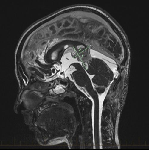
Magnetic Resonance Imaging ( MRI ) Scan
Pineoblastomas tend to appear as sizable (>4 cm) irregular masses often with evidence of invasion into adjacent brain 6,9.
Typical signal characteristics include :
T1: isointense to hypointense to adjacent brain
T2 isointense to adjacent brain areas of cyst formation or necrosis may be present
T1 C+ (Gd): vivid heterogenous enhancement
DWI/ADC restricted diffusion due to dense cellular packing
ADC values are typically ~400-800 mm2/s ADC values can aid in differentiation of pineoblastoma/PNET from germ cell tumors in a population of children with pineal masses 2).
Central necrosis is sometimes present which can make the mass appear centrally cystic and thus can roughly mimic a pineal cyst, although the latter should have a smooth thin wall
Screening of the whole neural axis is necessary as CSF seeding is seen in 45% of cases.
Differential diagnosis
General imaging differential considerations include:
other pineal parenchymal tumours
pineocytoma: mature well-differentiated tumor: smaller and better circumscribed
pineal parenchymal tumour with intermediate differentiation
papillary tumour of the pineal region
germ cell tumours
germinoma
marked male predominance engulfed calcification ADC values are typically much higher (~1000-2000 mm2/s 7)
embryonal carcinoma
choriocarcinoma
teratoma: may contain fat
pineal cyst thin (<2mm) wall
astrocytoma of pineal gland
metastasis
medulloblastoma
Imaging is very similar located in the vermis rather than pineal region but can be difficult to distinguish if very high in the vermis and very large
Treatment
Treatment is usually a combination of surgery including Stereotactic Image guided Biopsy, VP shunting or endoscopic third ventriculostomy -for hydrocephalus; Microsurgery for tumor removal, chemotherapy and radiation including Stereotactic Radiotherapy and Proton therapy stated Dr Prem Pillay , a Neurosurgeon who specializes in treating Pineal tumors including Pineoblastoma (He is currently Medical Director of the Singapore Brain Spine Nerves Center and was previously Chief Resident in Neurosurgery at the Cleveland Clinic, USA and fellow at MD Anderson Cancer Center and the Hospital for Sick Children, Toronto).
Outcome
They are the most aggressive and highest grade tumour among pineal parenchymal tumours.
Prone to CSF seeding, which is present in 15% of patients at the time of diagnosis.
Aggressive tumor resection was associated with improved survival only in older pediatric patients. Radiotherapy was more effective in patients receiving surgery. Age-stratified approaches might allow for improved disease management of pediatric pineoblastoma 3).
The role of surgery and adjuvant radiotherapy on overall survival remains to be defined 4).
Case series
A total of 211 pediatric (age 0-17 yr) histologically confirmed pineoblastoma patients diagnosed between 2004 and 2015 were queried from the National Cancer Database. Wilcoxon rank-sum statistics and chi-squared analyses were used to compare continuous and categorical variables, respectively. Univariable and multivariable Cox regressions were used to evaluate the prognostic impact of covariates. Propensity-score matching was used to balance baseline characteristics.
Older patients (age ≥ 4 yr) experienced improved overall survival compared to younger patients (age < 4 yr) (hazard ratio [HR] = 0.41; 95% CI 0.25-0.66). Older patients (adjusted odds ratio [aOR] = 5.21; 95% CI 2.61-10.78) and those residing in high-income regions (aOR = 3.16; 95% CI 1.21-8.61) received radiotherapy more frequently. Radiotherapy was independently associated with improved survival in older (adjusted HR [aHR] = 0.31; 95% CI 0.12-0.87) but not younger (aHR = 0.64; 95% CI 0.20-1.90) patients. The benefits of radiotherapy were more pronounced in patients receiving surgery than in those not receiving surgery (aHR [surgical patients] = 0.23; 95% CI 0.08-0.65; aHR [nonsurgical patients] = 0.46; 95% CI 0.22-0.97). Older patients experienced improved outcomes associated with aggressive resection (P = .041); extent of resection was not associated with survival in younger patients (P = .880).
Aggressive tumor resection was associated with improved survival only in older pediatric patients. Radiotherapy was more effective in patients receiving surgery. Age-stratified approaches might allow for improved disease management of pediatric pineoblastoma 5).
Using the Surveillance, Epidemiology, and End Results (SEER) cancer registry, Selvanathan et al., investigated clinical and pathological factors associated with outcome in paediatric pineoblastomas. Paediatric patients (< 16 years old) with pineoblastomas diagnosed between 1990 and 2007 were identified from the SEER database. Kaplan-Meier survival analysis and Cox models were used to examine the effect of variables on overall survival. The variables analysed included patient’s age at diagnosis, gender, race, tumour spread and size, surgical resection and the use of adjuvant radiotherapy.
Seventy-eight patients were identified from the database. Twelve patients were excluded as 11 had no surgery and one patient was excluded as the surgical status was unknown. Analysis of the remaining 66 patients revealed a median age at diagnosis of 5.5 years. Three patients underwent biopsy. Seventeen patients underwent full and partial resection, respectively. A further 46 patients underwent surgery the nature of which was not recorded. Thirty-nine patients (59.1%) received adjuvant radiotherapy. Eight patients (12.1%) had both surgery (full or partial resection) and radiotherapy. The median overall survival was 40.5 months. Univariate analysis demonstrated that older age at diagnosis was the only positive predictor of overall survival.
This study represents the largest analysis of paediatric pineoblastomas to date. The only clinically relevant prognostic factor was older age at diagnosis. The role of surgery and adjuvant radiotherapy on overall survival remains to be defined 6).
Conclusion
Pineoblastoma, though rare and aggressive, can be managed effectively with early diagnosis and a tailored treatment plan. At Singapore Brain Spine Nerves Center, we are dedicated to providing expert, compassionate care for patients facing complex conditions like pineoblastoma. If you or a loved one are experiencing concerning neurological symptoms, seek professional medical evaluation promptly. Early intervention is crucial to achieving the best possible outcomes. Visit the Singapore Brain Spine Nerves Center for comprehensive support and personalised treatments.
References
1) , 4) , 6)
Selvanathan SK, Richards O, Alli S, Elliott M, Tyagi AK, Chumas PD. Outcome and prognostic features in paediatric pineoblastomas: analysis of cases from the Surveillance, Epidemiology, and End Results registry (1990-2007). Acta Neurochir (Wien). 2019 May 18. doi: 10.1007/s00701-019-03909-1. [Epub ahead of print] PubMed PMID: 31104125.
2)
Choudhri AF, Whitehead MT, Siddiqui A, Klimo P Jr, Boop FA. Diffusion characteristics of pediatric pineal tumors. Neuroradiol J. 2015 Apr;28(2):209-16. doi: 10.1177/1971400915581741. Epub 2015 May 11. PubMed PMID: 25963154.
3) , 5)
Jin MC, Prolo LM, Wu A, Azad TD, Shi S, Rodrigues AJ, Soltys SG, Pollom EL, Li G, Hiniker SM, Grant GA. Patterns of Care and Age-Specific Impact of Extent of Resection and Adjuvant Radiotherapy in Pediatric Pineoblastoma. Neurosurgery. 2020 Feb 28. pii: nyaa023. doi: 10.1093/neuros/nyaa023. [Epub ahead of print] PubMed PMID: 32110805.


